Use Low Voltage FE-SEM to Overcome
Challenges of Imaging Biological Specimens Introduction
Biological samples span an incredible range of size scales, composition, structures, and morphology. Over the last few decades two complementary microscopy techniques for biological specimens have moved to the forefront, confocal laser scanning microscopy and scanning electron microscopy (SEM). With respect to the SEM many biological specimens share a similar set of issues, namely low conductivity of the sample and the typical glass substrates.Generally samples examined in the SEM need to be electrically conducting
in order to minimize charge buildup on the sample from the electron beam. Charge buildup can severely degrade the resultant image data. Advances in SEM to address a wider range of samples have led to brighter sources, fi eld emission fi laments, low vacuum also termed environmental SEM (eSEM), and low voltage SEM (LV-SEM). Three approaches can be employed to minimize charging. One approach is to metal coat the sample with an inert metal like gold. Another option is to increase the pressure in the sample chamber (eSEM) so that the gas molecules balance the charge. The third option is to decrease the electron beam voltage (LV-SEM) so
that the beam energy is at the charge equilibrium point.Biological samples typically have low conductivity and fragile structures and
therefore are subject to charge buildup and electron beam damage. One way to mitigate both of these issues is to use LV-SEM. However, low beam voltage operation of the SEM normally results in low resolution images. In order to improve resolution and
contrast in the SEM, increasing the source brightness and decreasing the initial probe size by using a field emission filament is a good solution. Agilent’s 8500 FE-SEM is a low voltage, fi eld emission SEM which employs a novel electrostatic lens design. This innovative design allows for high resolution imaging of biological samples, typically without the need for metal coating. The 8500 FE-SEM was used to image the following types of biological specimens, diatoms, human hair, mouse small intestine, cells, and bacteria.
Diatoms
Diatoms are a major group of algae and are one of the most common types of phytoplankton. Most diatoms are unicellular, although they can exist as colonies (Fragillaria, Meridion, Tabellaria, or Asterionella). A characteristic feature of diatom cells is that they are encased within a unique cell wall made of silicates called a frustule. These frustules show a wide diversity in form, but usually consist of two asymmetrical sides with a split between them.
Fossil evidence suggests that they originated during, or before, the early Jurassic Period. Diatom communities are a popular tool for monitoring environmental conditions, past and present, and are commonly used in studies of water quality.
The bio-mineralization of silica by diatoms may also prove to be useful for nanotechnology. Diatom cells repeatedly and reliably manufacture valves and pores of various shapes and sizes. This could potentially allow diatoms to manufacture nano-scale structures which may be of use in a range of devices. Using an appropriate artificial selection procedure, diatoms that produce valves of particular shapes and sizes could be engineered in the laboratory, and then used to mass produce nano-scale components.
LV FE-SEM was used to image diatoms prepared on SEM conductive carbon adhesive tape, see Figures 1-3. With 1 kV accelerating voltage the diatom samples were imaged uncoated with no charge buildup issues.

Human Hair
The desire for products that improve the look and feel of hair has created a huge industry for hair care. Hair care technology has advanced the cleaning, protection, and restoration of desirable hair properties by altering the chemical and physical properties of the hair surface. Shampoo is used to clean hair and conditioner is used to coat the hair with a thin film in order to protect it and provide desirable look and feel.
The success of a cosmetic product is based not only on its properties, but also on the way it interacts with the biological specimen.

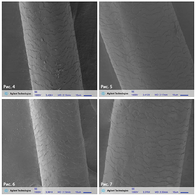
Most people who have both brown and grey hair types perceive definite differences between pigmented (Figures 4-5) and unpigmented (grey) hair (Figures 6-7). The perceived differences are effectively mechanical: grey hair is said to be "stiffer," more "wiry," and generally more unruly. For one test subject, grey hair was found to be stiffer than brown hair [2] because the diameter was larger by 36%. These results are consistent with previous studies, leading to the suspicion that hair products aimed at changing the composition of grey hair are unlikely to be effective in changing stiffness.[2]
Mouse Intestine
intestine is part of the digestive system where food is broken down to its smallest component and nutrients absorbed. Mouse intestine is an animal model for understanding digestive disorders and infectious diseases. LV FE-SEM with 1 kV accelerating voltage was used to image mouse intestine microvilli tissue samples, see Figures 8-10. In Figure 10 cross-sections of several microvilli can be seen.
Fixed Cells
The identification and characterization of cell substructures at high resolution requires the use of electron microscopy techniques. A wide range of magnifications is possible, from 10 times (about equivalent to that of a powerful hand-lens) to more than 500,000 times, about 250 times the magnification limit of the best light microscopes.
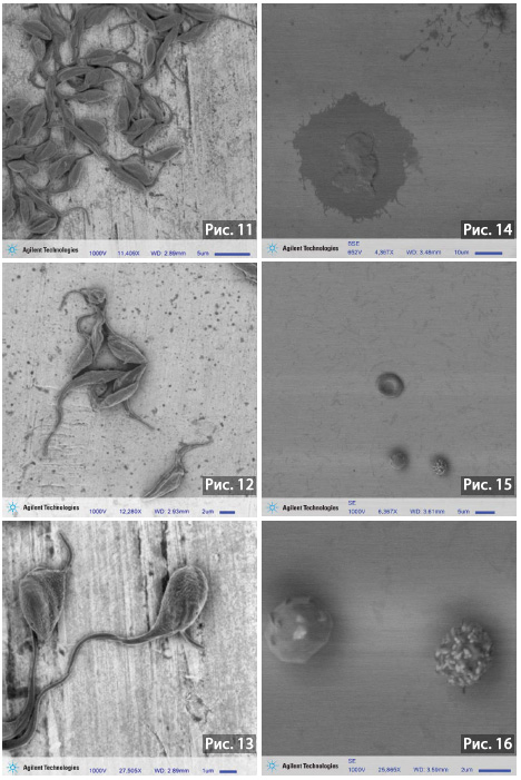
SEM allows the direct visualization of macromolecular structures, cilia, flagella, and cell surface morphology. Due to the very narrow electron beam, SEM micrographs have a large depth of field yielding a characteristic threedimensional appearance useful for understanding the surface structure of a sample. However, the trade-off with optical microscopy is that SEM generates only static images of fixed samples and therefore cannot give unambiguous information about cellular and protein dynamics. The investigation of active processes and analysis of protein dynamics has greatly benefited from the development of molecular biology techniques whereby specific proteins tagged with a fluorescent moiety for real-time optical microscopy visualization. Therefore LV FE-SEM is an excellent complementary technique for cell biologists, allowing the visualization and characterization of surface morphologies and substructures that are unattainable with optical microscopy. A variety of cell types were imaged without metal coating using LV FE-SEM with 1 kV accelerating voltage, see Figures 11-16.

Bacteria
Bacillus megaterium is a Gram-positive, rod-shaped, endospore forming species of bacteria that is used as a soil inoculant in agriculture. The bacterium is arranged into a chain-link structure like streptobacillus. Bacillus megaterium is one of the largest eubacteria found in soil. Groups of the bacteria are often found in chains where the cells are joined together by polysaccharides on the cell walls. Bacillus megaterium is able to survive in some extreme conditions such as desert environments due to the spores it forms. Bacillus megaterium produces penicillin amidase used for making penicillin. It also can produce enzymes for modifying corticosteroids, as well as several amino acid dehydrogenases.

Bacillus megaterium is a good source for industrial produced proteins because it is both a desirable cloning host and produces a large variation of enzymes. This species is good cloning host because it is able to house numerous plasmid vectors while remaining stable due to its unique external proteases. For example, many synthetic penicllins have been derived using the penicillin amidase in the bacteria and harvested glucose dehydrogenase is used in glucose blood tests. The biotechnological applications of the Bacillus megaterium provide an abundance of different proteins that are used in important medical, scientific and industrial advances. LV FE-SEM with 1 kV accelerating voltage was used to image Bacillus megaterium, see Figures 17-19.
Engineered Cell Substrates
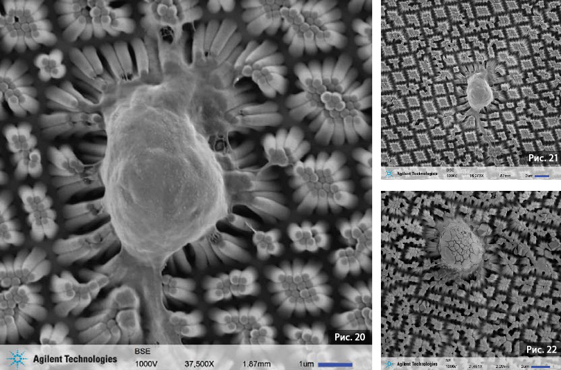
Current in vitro cell culture technologies present some limitations as they can't simulate in vivo situations. In reality, in vivo cells function in a three-dimensional structure, or tissue, where they have a close contact with adjacent cells. Cell growth and viability is enhanced by increased oxygenation and better modulus match of the PDMS (polydimethylsiloxane) growth surface (Figures 20-22). Commonly cells on PDMS require metal coating for SEM imaging, see Figure 20. However, the metal coating obscures the cell surface morphology. By using LV FE-SEM it is possible to image these specimens with no metal coating, see Figure 21-22. By imaging without metal coating, the cell surface morphology is clearly visible.

Conclusions
LV FE-SEM provides a straightforward technique for high resolution imaging of fixed biological samples, typically without the need for metal coating. Although the biological samples span an incredible range of size scales, composition, and structures, the morphological features of interest could easily be investigated with the Agilent 8500 FE-SEM.

